The Role of Desmin Variants In Familial Atrial Fibrillation
Atrial fibrillation (AF) is the most common sustained cardiac arrhythmia in the Western world. AF is closely correlated with traditional risk factors. Traditional risk factors include lifestyle related hypertension, diabetes and obesity. Researchers increasingly recognise that a genetic component may underlie AF development. Recent study findings indicate that a several gene variants are associated with AF. These gene variants include genes encoding sodium, potassium and calcium channels. In addition, patients may also carry variations in genes encoding the cytoskeletal proteins and some variations are related to AF development.
Cytoskeletal proteins are proteins which provide cell structure and cell contraction and as such ensures optimal cell and organ function. Gene variants in cytoskeletal proteins that associate with AF include:
- lamin A/C
- desmin
- desmoplakin
Patients carrying gene variants in cytoskeletal genes may develop AF at a young age. But how do these gene variants trigger AF? What are the underlying molecular mechanisms?
PhD student Wei Su of the research team of Prof. Bianca Brundel, is studying how desmin variants may cause AF. Hereto, we first need to understand the physiological role of desmin in a healthy heart. This knowledge may elucidate leads for further research in AF.
Desmin is needed for normal heart function
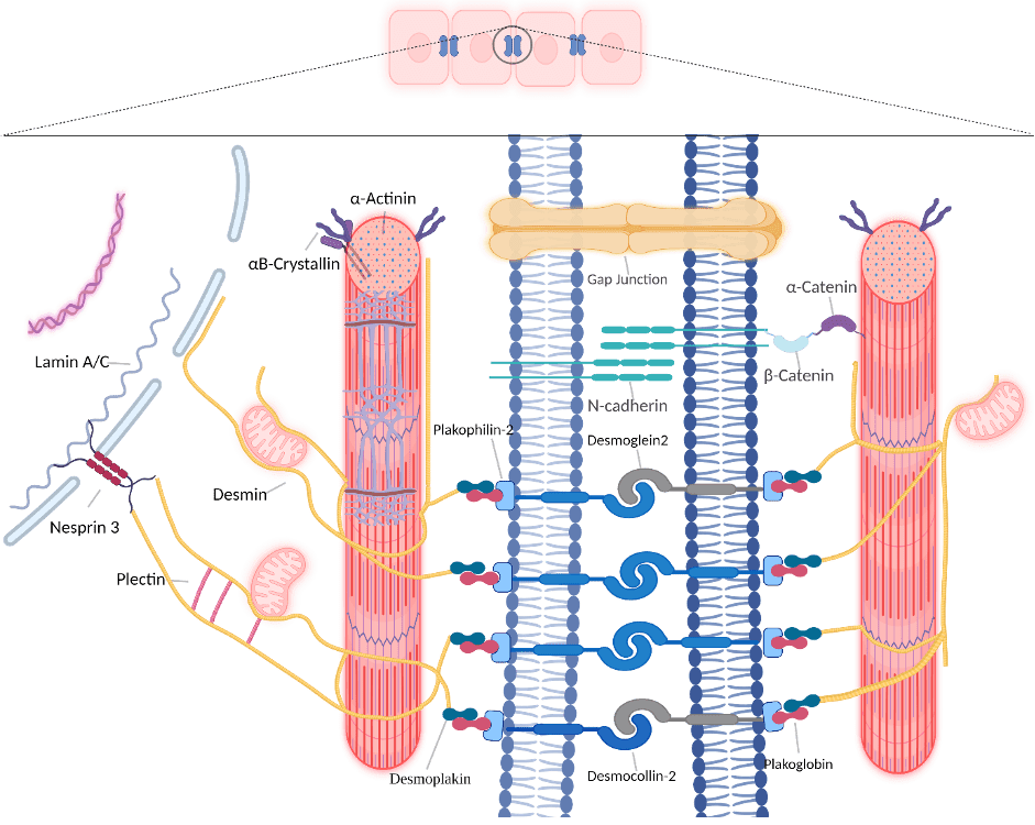
Desmin is a classical type III intermediate filament protein encoded by the DES gene. Intermediate filament proteins are proteins that interlink cardiomyocytes in the atria. Also, within the cardiomyocyte itself, intermediate filament proteins link cell organelles and enable crosstalk within the cells. In addition, they provide cell structure (Figure 1).

Figure 1. Overview of desmin network (in yellow) in and between cardiomyocytes (adapted from Wei Su et al).
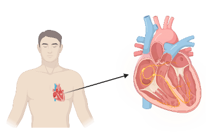
Desmin is synthesised in large quantifies in cardiac, skeletal, and smooth muscle cells. In the heart, desmin is widely expressed in (Figure 2):
- the atrioventricular node; this node is primarily an electrical gatekeeper between the atria and ventricles and introduces a delay between atrial and ventricular excitation, allowing for efficient ventricular filling
- the atrioventricular bundle; a bundle of specialized heart muscle fibers that conducts impulses from the atrioventricular node to the ventricles.
- ventricular trabeculations: protrusions from the developing ventricular wall into the ventricular lumen.

Figure 2. Expression pattern (yellow) of desmin in the conduction system of the heart
As these specific desmin locations are all involved in the cardiac conduction system, it is not a surprise that variants in desmin will result in cardiac arrhythmias including AF.
Desmin function and molecular interaction partners in the atria
The important function of desmin in the cardiac conduction system is related to the distinctive property of desmin to form intra- and intercellular networks by connecting and anchoring various cytoskeletal structures and organelles. The following examples are illustrated in figure 1:
- Desmosomes. These are are intercellular junctions that provide strong adhesion between cells.
- Mitochondria. These are organelles that produce energy.
- Nuclei that contain DNA.
- Costameres. These are proteines that connect the sarcomere of the muscle to the cell membrane.
- Z-bands. This is a protein structure that connects actin filaments of opposite polarity from adjacent sarcomeres (smallest contractile unit of the cardiomyocytes).
The desmin network plays an important role in striated myocardium development, maintenance and consequently electrical and contractile function of the atria. Hereto, desmin integrates and coordinates most cellular components necessary for proper mechanochemical signaling, organelle cross-talk, energy production, and trafficking processes required for proper atrial tissue homeostasis.
Desmin variants, atrial fibrillation and other clinical arrhythmias
Because desmin is located in various human tissues, the clinical phenotypes associated with DES variants are diverse. So far, over 60 different pathogenic DES variants have been described. Most patients with DES variants develop combined skeletal and cardiac diseases (myopathies). Also, most DES variants related to electrical conduction defects and arrhythmias, are missense mutations, often located in coil 2B or near the carboxyl terminus of desmin (Figure 3). Eight DES variants are related with AF onset at a young age.

Figure 3. Schematic overview of cardiac conduction defects and arrhythmias associated with DES variants (adapted from Wei Su et al).
Potential mechanism how desmin variants cause atrial fibrillation
Within the DnAFix project, our research team started to investigate the molecular root causes of DES variant-induced AF. Here are some of the key findings:
- Desmin aggregation within cardiomyocytes is the most significant histopathological hallmark of desmin cardiomyopathies. These desmin aggregates may lead to cardiomyopathy onset.
- In an electrocardiography study, desmin deficient mice show a significant reduction in atrial and in the meantime a prolonged ventricular refractory period, indicating susceptibility for AF onset.
- Mitochondrial function and structure abnormalities seem to be the earliest detected defects in cardiomyocytes lacking desmin. Mitochondrial abnormalities were also found in to play a role in clinical AF.
- Moreover, knockdown of desmin in cardiomyocytes results in an abnormal distribution of Ca2+. A marked increase in cytosolic Ca2+ concentration and a decrease in the sarcoplasmic reticulum Ca2+ concentration was found. As Ca2+ plays a key role in the contractile function of cardiomyocytes, and cytosolic Ca2+ overload is a trigger for AF, DES variants may alter the Ca2+ handling and trigger AF.
Towards future therapies for atrial fibrillation induced by desmin variants
One of the critical steps for a future therapeutic approach for desmin cardiomyopathies is to inhibit desmin aggregation in the cardiomyocytes. As a previous experimental study showed, the non-toxic heat shock protein inducer geranylgeranylacetone (GGA) inhibits desmin-related cardiomyopathy. GGA is an interesting compound to be tested in future clinical studies as 3 days of oral GGA treatment induces heat shock protein levels in atria of patients undergoing elective cardiac surgery. Of note, GGA also protects against experimental AF onset and may help to recover from AF-induced damage.
Modulating mitochondrial function, with nutraceuticals such as nicotinamide, or mitochondria-targeted peptides, gene, and stem cell therapy or muscle-specific gene transfer approaches are active areas of research that promise effective treatments for future treatment of AF.
In summary
The desmin network plays a significant role in maintaining the structural and mechanical integrity of (atrial) cardiomyoctyes. Moreover, desmin is involved in cardiomyocyte function by modulating cellular signaling, contraction and calcium homeostasis. As desmin is highly expressed in structures of the cardiac conduction system, the majority of pathogenic DES variants cause cardiac arrhythmia, cardiac conduction defects as well as atrial fibrillation. On the molecular level, impaired desmin causes severe cytoskeletal defects, desmin aggregation, and abnormal distribution of Ca2+, that collectively may drive cardiac arrhythmias such as atrial fibrillation.
The complete research article entitled ‘Desmin variants: Trigger for cardiac arrhythmias?’ is written by Wei Su, Stan van Wijk and Bianca Brundel and published in Front Cell Dev Biol. 2022 Sep 9;10:986718. doi: 10.3389/fcell.2022.986718. eCollection 2022.
Do you know someone who might be interested in this?


